WesternU pilot tests Anatomage Navigator
Western University of Health Sciences is the first university to utilize the Anatomage Navigator, a volumetric inner anatomy visualization tool with tracking stylus.
The Anatomage Navigator adds another dimension to WesternU’s J and K Virtual Reality Learning Center (VRLC), which includes the Anatomage Virtual Dissection Table. The dissection table projects three-dimensional images constructed from a combination of computerized tomography (CT) scans of the human body, magnetic resonance imaging (MRI), X-rays and digital photographs of a cadaver frozen in a block of blue gel. The digital files are integrated together into three-dimensional models that allow users to manipulate the digital files in multiple ways.
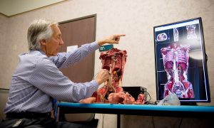
The Navigator includes a plaster torso model created with three-dimensional color printing technology, along with a high-definition display that projects multiple cross-sectional views. The stylus tracker allows the user to explore real anatomy without the need for cadavers.
“We can remove and hold some of the organs that are molded and cast in silicone, similar to the texture and weight of actual organs,” said College of Dental Medicine Associate Dean for Simulation, Immersion and Digital Learning Robert Hasel, DDS. “For example, the liver is made with a semi-transparent material that shows the lobes of liver, the vasculature and the ducts. The lungs are made out of light and spongy material. As you hold the lung in your hand, it builds a 3-D mental relationship in your mind to augment what you’ve seen on the Anatomage table and the iPad or textbook.
“By placing the stylus probe on certain structures, you can see how they relate to surrounding structures,” Dr. Hasel added. “For instance, you can look at which ribs are behind the lung tissue and where the kidneys are located in relation to the diaphragm, and at the same time see the cross-sections in three different planes.”
The Navigator is the latest technology to arrive at the VRLC, which features an Anatomage Virtual Dissection Table, two zSpace 4D displays, two Oculus Rifts, eight iPad stations and fusion.tech’s Stanford anatomical models.
“We’re the showcase for Anatomage technology for North America,” Hasel said. “We are piloting the Navigator and testing it out to provide feedback on construction and design and to develop curriculum for it.”
WesternU is one of the first pilot programs to start integrating the Navigator into its anatomy curriculum. College students will start utilizing the Navigator during an anatomy workshop in June.
The Navigator allows users to load in and view their own CT/MRI contents. These three-dimensional volumes can be mapped to generic 3-D print models or ones made from that specific patient’s anatomy. For students, this reinforces the comprehension of 3-D spatial relationships and visualization of anatomy in their individual patients, said Kris Thomson, Anatomage Director of Table Applications.
“We’re excited to see how the Navigator can be integrated into the curriculum,” Thomson said. “From an educational point of view, not everyone is able to comprehend anatomy in the same way. Sometimes you need that physical model. We feel a realistic 3-D print combined with software can really help with that.”