WesternU CPM and CETL develop 3D animations depicting lower extremity biomechanics and foot and ankle surgical procedures
Western University of Health Sciences College of Podiatric Medicine (CPM) students have cutting-edge 3D animations and models available at their fingertips thanks to a collaboration between CPM Professor Stephen Wan, DPM, FACFAS, CPM Assistant Professor Saba Sadra, DPM, and Gary Wisser of WesternU’s Center for Excellence in Teaching & Learning (CETL).
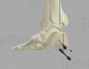
The animations and models depict lower extremity biomechanics as well as foot and ankle surgical procedures to improve the educational experience for CPM students. This was in response to students having a difficult time understanding these topics because they could not “visualize” the process, Sadra said.
“The primary objectives of our project were to enhance the understanding of foot and ankle joint movements, normal and abnormal ontogeny, gait patterns, and surgical procedures among students. By developing intricate 3D models and animations, our team aimed to provide a comprehensive and immersive learning experience,” Sadra said. “These animations allow students to visualize the axis of motion for foot and ankle joints, observe normal gait in a detailed gait model, and comprehend the intricacies of lower extremity development and surgical procedures.”
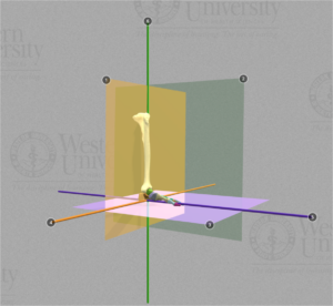
Prior to the incorporation of these 3D animations into the curriculum, students traditionally relied on static images, textbooks, and lectures, which often presented limitations in conveying the dynamic and three-dimensional nature of these complex topics.
“Our project offers an interactive and a visually engaging platform, allowing students to explore and understand foot and ankle biomechanics and surgical procedures in a more profound and practical manner,” Sadra said.
The project took about two-and-a-half years from conception to final animation, Wisser said.
“Early on we agreed this project would only be worth doing if we stayed dedicated to creating a quality product that would stand the test of time,” he said. “We started with a high-definition model of foot bones that I created about a year earlier. I scanned individual foot and leg bones in high resolution that showed all of the surface details for each bone.”
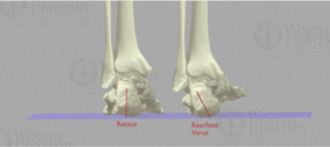
Drs. Sadra and Wan are the content experts for the project. They worked meticulously to make sure the bone positions were correct in both weight-bearing and non-weight bearing motions, Wisser said.
“Three things that really make this project unique are the quality of the models, the dedication Dr. Sadra and Dr. Wan gave to the accuracy of positions and motions, and the fact that the end user has total control of the position of viewing,” Wisser said. “The animations can be zoomed in and rotated into any position desired.”
The trio started this project during the COVID-19 pandemic. Every week, Dr. Wan drove to Wisser’s house and Dr. Sadra joined via Zoom, and they would spend a few hours making sure the anatomical positions and medical procedures were as close to perfect as they could make them, Wisser said. They needed to look correct from every angle.
“That dedication to accuracy by two medical professionals is what helped raise the bar for this kind of illustration,” Wisser said.
