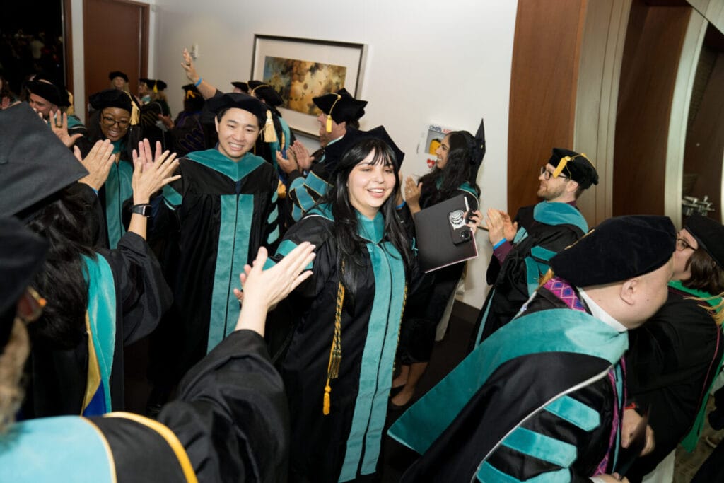
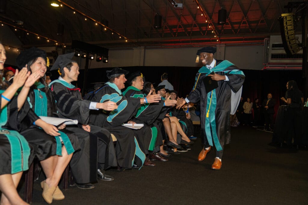
Western University of Health Sciences’ College of Optometry held its Commencement ceremony on May 13, 2025 in Pomona, California. College of Optometry graduate speaker Gerard Joseph Sim, OD ’25, said his classmates demonstrated humanism throughout their time at WesternU, carving time out of their busy schedules to volunteer in the community and abroad in Mexico and Fiji.
However, humanism is not only compassion toward our patients, but also the sense of support and community we provide for each other, Sim said. This has been exemplified by their mentors, families and amongst each other.
“It’s the friendships that we shared with each other, whether it was the first friend we met during orientation or the bonds that we created within our clinic groups, the friendships we created will carry us forward professionally. One of my favorite memories working as one of your lab monitors, besides cleaning up after you all, is getting to see you all practice. I love seeing you encourage your friend or your lab partner, providing great feedback and practicing for long hours into the night until you prepared each other for your proficiencies,” Sim said. “No matter where the next adventure takes us, I have faith in the support we will give to each other. As we embark on the next chapter of our lives, I know we are ready. We have developed the resilience to overcome any obstacle, the knowledge to improve our patients’ quality of life, and this wonderful community of support that we created here at WesternU.”
Commencement feels like any other day, said College of Optometry graduate Elia Rptchian, OD ’25, then added, “but it feels like my day and that it’s the start of the next journey. I got experiences that I never thought I would get and I did things I never thought I would do myself. I surprised myself along the way, which was nice.”
“There was a lot of hard work and a lot of sleepless nights and tears leading up to this moment,” said College of Optometry graduate Michelle Cristi, OD ’25. “It makes me proud looking back on that experience.”
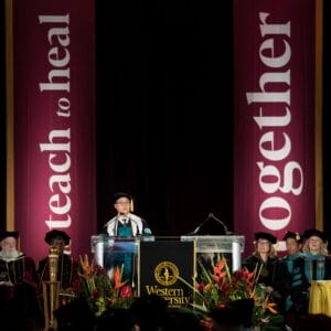
Optometry is about more than eyesight, it’s about seeing the whole person, said WesternU Provost and Chief Academic Officer Paula M. Crone, DO ’92. It’s about restoring confidence, enhancing independence and opening doors to opportunity for your patients.
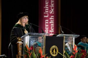
“As graduates of WesternU, you are uniquely prepared to practice with skill, heart, and with integrity. As doctors of optometry, you are entering a profession that does more than correct vision.
It changes lives. It’s a vital part of our health care system, one that promotes wellness, enhances learning and productivity, detects systemic disease and, for many, restores a sense of independence and possibility,” Crone said. “Your education has prepared you to deliver expert care, and to lead with both clinical skill and human connection. But beyond what you have learned, what will define your life is how you care. how you build trust, listen deeply, and make your patients feel seen, valued and understood. As you take this next step, remember that your education doesn’t end here. It evolves. Stay curious, lead with empathy, and never underestimate the impact of helping someone see the world more clearly.”